150 Years of Woods Hole Science
30
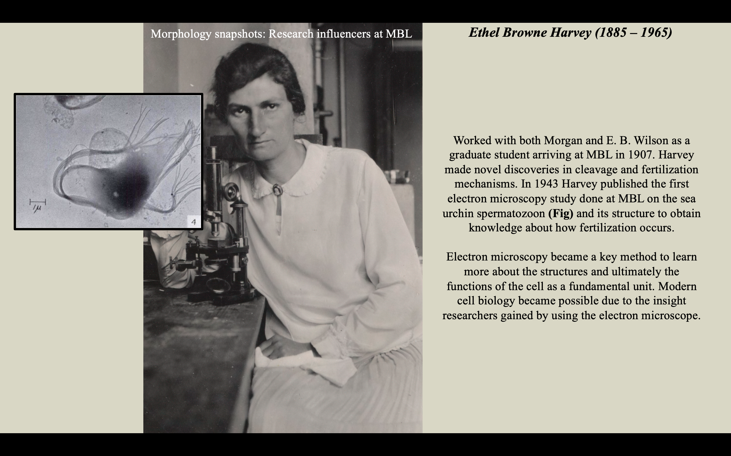
Fig: Plate II, Figure 4 from Harvey & Anderson, 1943. Harvey and Anderson’s electron microscope image of the A. punctulata spermatozoon showing remains of the tail and membrane following rupture by a change in tonicity.
Background image: Ethel Browne Harvey, Marine Biological Laboratory Archives, from https://hdl.handle.net/1912/16775
References:
Harvey, E.B. and Anderson, T.F., 1943. The spermatozoon and fertilization membrane of Arbacia punctulata as shown by the electron microscope. The Biological Bulletin, 85(2), pp.151-156. – Fig.